The third stage of evolution is the appearance of the cell.
Molecules of proteins and nucleic acids (DNA and RNA) form a biological cell, the smallest unit of life. Biological cells are the "building blocks" of all living organisms and contain all the material codes of development.
For a long time, scientists considered the structure of the cell to be extremely simple. The Soviet Encyclopedic Dictionary interprets the concept of a cell as follows: "A cell is an elementary living system, the basis of the structure and life of all animals and plants." It should be noted that the term "elementary" in no way means "the simplest". On the contrary, a cell is a unique fractal creation of God, striking in its complexity and, at the same time, in the exceptional coherence of the work of all its elements.
When we managed to look inside with the help of an electron microscope, it turned out that the device of the simplest cell is as complex and incomprehensible as the Universe itself. Today it has already been established that "A cell is a special matter of the Universe, a special matter of the Cosmos." One single cell contains information that can be put into only a few tens of thousands of volumes of the Great Soviet Encyclopedia. Those. the cell, among other things, is a huge "bioreservoir" of information.
The author of the modern theory of molecular evolution, Manfred Eigen, writes: “In order for a protein molecule to form by chance, nature would have to do about 10130 trials and spend on this such a number of molecules that would be enough for 1027 Universes. If the protein was built intelligently, that is, that the validity of each move could be checked by some selection mechanism, it took only about 2000 attempts. We come to a paradoxical conclusion: the program for building a "primitive living cell" is encoded somewhere at the level of elementary particles ".
And how could it be otherwise. Each cell, having DNA, is endowed with consciousness, is aware of itself and other cells, and is in contact with the Universe, being, in fact, a part of it. And although the number and variety of cells in the human body is amazing (about 70 trillion), they are all self-similar, just as all processes occurring in cells are self-similar. In the words of the German scientist Roland Glaser, the design of biological cells is "very well thought out." Who is well thought out?
The answer is simple: proteins, nucleic acids, living cells and all biological systems are the product of the creative activity of the intellectual Creator.
What is interesting: at the atomic level, there are no differences between the chemical composition of the organic and inorganic world. In other words, at the level of an atom, a cell is created from the same elements as inanimate nature. Differences are found at the molecular level. In living bodies, along with inorganic substances and water, there are also proteins, carbohydrates, fats, nucleic acids, the ATP synthase enzyme, and other low molecular weight organic compounds.
To date, the cell has been literally dismantled into atoms for the purpose of study. However, it is not possible to create at least one living cell, because to create a cell means to create a particle of the living Universe. Academician V.P. Kaznacheev believes that "a cell is a cosmic-planetary organism... Human cells are certain systems of ether-torsion biocolliders. In these biocolliders, processes unknown to us occur, materialization of cosmic forms of flows takes place, their cosmic transformation takes place, and due to this particles materialize" .
Water.
Almost 80% of the cell mass is water. According to Doctor of Biology S. Zenin, water, due to its cluster structure, is an information matrix for managing biochemical processes. In addition, it is water that is the primary "target" with which sound frequency oscillations interact. The orderliness of cellular water is so high (close to the orderliness of a crystal) that it is called a liquid crystal.
Squirrels.
Proteins play an important role in biological life. The cell contains several thousand proteins that are unique to this type of cell (with the exception of stem cells). The ability to synthesize its own proteins is inherited from cell to cell and persists throughout life. During the life of the cell, proteins gradually change their structure, their function is impaired. These spent proteins are removed from the cell and replaced with new ones, thanks to which the vital activity of the cell is preserved.
First of all, we note the building function of proteins, because they are the building material that makes up the membranes of cells and cellular organelles, the walls of blood vessels, tendons, cartilage, etc.
The signaling function of proteins is extremely interesting. It turns out that proteins are able to serve as signaling substances, transmitting signals between tissues, cells or organisms. The signaling function is performed by hormone proteins. Cells can communicate with each other at a distance using signaling proteins transmitted through the intercellular substance.
Proteins also have a motor function. All types of movement that cells are capable of, such as muscle contraction, are performed by special contractile proteins. Proteins also perform a transport function. They are able to attach various substances and transfer them from one place in the cell to another. For example, the blood protein hemoglobin attaches oxygen and carries it to all tissues and organs of the body. In addition, proteins also have a protective function. When foreign proteins or cells are introduced into the body, special proteins are produced in it that bind and neutralize foreign cells and substances. And finally, the energy function of proteins is that with the complete breakdown of 1 g of protein, energy is released in the amount of 17.6 kJ.
Cell structure.
The cell consists of three inextricably linked parts: membrane, cytoplasm and nucleus, and the structure and function of the nucleus in different periods of the cell's life are different. For the life of a cell includes two periods: division, as a result of which two daughter cells are formed, and the period between divisions, which is called interphase.
The cell membrane directly interacts with the external environment and interacts with neighboring cells. It consists of an outer layer and a plasma membrane located underneath. The surface layer of animal cells is called the glycocalys. It connects cells with the external environment and with all the substances surrounding it. Its thickness is less than 1 micron.
Cell structure
The cell membrane is a very important part of the cell. It holds together all cellular components and delimits the external and internal environment.
There is a constant exchange of substances between cells and the external environment. Water, various salts in the form of individual ions, inorganic and organic molecules enter the cell from the external environment. Metabolism products, as well as substances synthesized in the cell: proteins, carbohydrates, hormones, which are produced in the cells of various glands, are excreted into the external environment through the membrane from the cell. The transport of substances is one of the main functions of the plasma membrane.
Cytoplasm- an internal semi-liquid medium in which the main metabolic processes take place. Recent studies have shown that the cytoplasm is not a kind of solution, the components of which interact with each other in random collisions. It can be compared to jelly, which begins to "tremble" in response to external influences. This is how the cytoplasm perceives and transmits information.
The nucleus and various organelles are located in the cytoplasm, which are united by it into one whole, which ensures their interaction and the activity of the cell as a single integral system. The nucleus is located in the central part of the cytoplasm. The entire inner zone of the cytoplasm is filled with the endoplasmic reticulum, which is a cellular organoid: a system of tubules, vesicles and "cisterns" delimited by membranes. The endoplasmic reticulum is involved in metabolic processes, providing the transport of substances from the environment to the cytoplasm and between individual intracellular structures, but its main function is participation in protein synthesis, which is carried out in ribosomes. - microscopic little bodies of round shape with a diameter of 15-20 nm. The synthesized proteins are first accumulated in the channels and cavities of the endoplasmic reticulum and then transported to the organelles and cell sites where they are consumed.
In addition to proteins, the cytoplasm also contains mitochondria, small bodies 0.2-7 microns in size, which are called the "power stations" of cells. Redox reactions occur in mitochondria, providing cells with energy. The number of mitochondria in one cell varies from a few to several thousand.
Nucleus- the vital part of the cell, controls the synthesis of proteins and through them all the physiological processes in the cell. In the nucleus of a non-dividing cell, the nuclear membrane, nuclear juice, nucleolus and chromosomes are distinguished. Through the nuclear envelope, there is a continuous exchange of substances between the nucleus and the cytoplasm. Under the nuclear envelope - nuclear juice (semi-liquid substance), which contains the nucleolus and chromosomes. The nucleolus is a dense rounded body, the dimensions of which can vary widely, from 1 to 10 microns and more. It consists mainly of ribonucleoproteins; participates in the formation of ribosomes. Usually there are 1-3 nucleoli in a cell, sometimes up to several hundred. The nucleolus consists of RNA and protein.
With the advent of the cell, Life arose on Earth!
To be continued...
summary of other presentations"Methods of teaching biology" - School zoology. Introducing students to the use of scientific zoological data. Moral education. Additional consecration of the chicken coop. Choice of methods. Life processes. Aquarium fish. Food. Ecological education. Materiality of life processes. Negative results. Attention of students. Mandatory form. Looking at small animals. Goals and tasks of biology. Story.
"Problem learning in biology lessons" - Knowledge. New textbooks. The path to a solution. Problem. Seminars. What is a task. Albrecht Durer. Problem-based learning in biology lessons. Non-standard lessons. What is meant by problem-based learning. The quality of life. Biology as a subject. Question. Problem solving lesson. Decreased interest in the subject. Problem-laboratory classes.
"Critical thinking in biology lessons" - Technology of "critical thinking". Using the technology of "development of critical thinking". Table for the lesson. Motivation for learning. Ecosystems. The meaning of "development of critical thinking". Technology features. RKM technology. Lesson structure. Main directions. History of technology. Pedagogical technologies. technology rules. Biology assignments. Photosynthesis. Techniques used at different stages of the lesson.
"Biology lessons with an interactive whiteboard" - Electronic textbooks. Benefits for students. An interactive whiteboard helps convey information to each student. didactic tasks. Solving biological problems. Benefits of working with interactive whiteboards. Presentation work. Work on comparing objects. Moving objects. Use of spreadsheets. The use of an interactive whiteboard in the process of teaching schoolchildren. Benefits for teachers.
"System-activity approach in biology" - Questions of the seminar. activity method. Dryopithecus. Extraterrestrial way of human origin. Lysosomes. Chemical organization. Gymnosperms. Metabolism. Analyzers. System-activity approach in teaching biology. Chromosomes. Cytoplasm. Blindness. Ear length. Human classification. Mammalian skeleton. Ways of human evolution. Mitosis. surface complex. Problem question. Nucleus. Nuclear shell.
"Computer on Biology" - Joint activities of students. Families of angiosperms. Interactive learning. learning models. An example of a grading system. Instruction card questions. An example of an instruction card. Researchers. Microgroups. Interactive learning technologies. Carousel. Interactive learning technologies. Interactive approaches in biology lessons. Group form of work. Tasks for groups of "researchers".
The connection of an organism with the environment, from a physicochemical point of view, is an open system, that is, a system where biochemical processes are ongoing. The initial substances come from the environment, and the substances that are also continuously formed are taken out. The balance between the rate and concentration of products of multidirectional reactions in the body is conditional, imaginary, since the intake and removal of substances do not stop. Continuous connection with the environment and allows us to consider a living organism as an open system.
The Sun is the source of energy for all living cells. Plant cells capture the energy of sunlight with the help of chlorophyll, using it for assimilation reactions during photosynthesis. Cells of animals, fungi, bacteria use solar energy indirectly, when splitting organic substances synthesized by an earthly plant.
Part of the nutrients of the cell is broken down in the process of cellular respiration, thus supplying the energy necessary for various kinds of cellular activity. This process takes place in organelles called mitochondria. The mitochondrion consists of two membranes: the outer one, which separates the organelle from the cytoplasm, and the inner one, which forms numerous folds. The main product of respiration is ATP. It leaves the mitochondria and is used as an energy source for many chemical reactions in the cytoplasm and cell membrane. If oxygen is required for the implementation of cellular respiration, then respiration is called aerobic respiration, but if the reactions take place in the absence of oxygen, then one speaks of anaerobic respiration.
For any kind of work done in a cell, energy is used in a single form - in the form of energy from the phosphate bonds of ATP. ATP is a highly mobile compound. The formation of ATP occurs at the inner membrane of mitochondria. ATP is synthesized in all cells during respiration due to the energy of oxidation of carbohydrates, fats and other organic substances. In the cells of green plants, the main amount of ATP is synthesized in chloroplasts due to solar energy. In them, during photosynthesis, many times more ATP is produced than in mitochondria. ATP decomposes with the breaking of phosphorus-oxygen bonds and the release of energy. This occurs under the action of the ATPase enzyme in the process of ATP hydrolysis - the addition of water with the elimination of a phosphoric acid molecule. As a result, ATP is converted into ADP, and if two molecules of phosphoric acid are split off, then into AMP. The cleavage reaction of each gram-molecule of acid is accompanied by the release of 40 kJ. This is a very large energy yield, so the phosphorus-oxygen bonds of ATP are commonly called macroergic (high-energy).
The use of ATP in the reactions of plastic exchange is carried out by their conjugation with the hydrolysis of ATP. Molecules of various substances are charged with energy by attaching the phosphorus group released during hydrolysis from the ATP molecule, that is, by phosphorylation.
A feature of phosphate derivatives is that they cannot leave the cell, although their "discharged" forms freely pass through the membrane. Due to this, phosphorylated molecules remain in the cell until they are used in the appropriate reactions.
The reverse process of converting ADP to ATP occurs by attaching a phosphoric acid molecule to ADP, releasing water and absorbing a large amount of energy.
Thus, ATP is a universal and immediate source of energy for cell activity. This creates a single cellular fund of energy and makes it possible to redistribute and transport it from one part of the cell to another.
The transfer of a phosphate group plays an important role in chemical reactions such as the assembly of macromolecules from monomers. For example, amino acids can only be combined into peptides if they have been previously phosphorylated. Mechanical processes of contraction or movement, the transfer of a solute against a concentration gradient, and other processes are associated with the expenditure of energy stored in ATP.
The energy exchange process can be represented as follows. High-molecular organic substances in the cytoplasm are enzymatically, by hydrolysis, converted into simpler ones, of which they consist: proteins - into amino acids, poly- and disaccharides - into monosaccharides (+ glucose), fats into glycerol and fatty acids. Oxidative processes are absent, little energy is released, which is not used and goes into thermal form. Most cells use carbohydrates first. Polysaccharides (starch in plants and glycogen in animals) are hydrolyzed to glucose. Glucose oxidation occurs in three phases: glycolysis, oxidative decarboxylation (Krebs cycle - citric acid cycle) and oxidative phosphorylation (respiratory chain). Glycolysis, as a result of which one molecule of glucose is split into two molecules of pyruvic acid with the release of two molecules of ATP, takes place in the cytoplasm. In the absence of oxygen, pyruvic acid is converted to either ethanol (fermentation) or lactic acid (anaerobic respiration).
If glycolysis is carried out in animal cells, the six-carbon glucose molecule breaks down into two molecules of lactic acid. This process is multi-stage. It is carried out sequentially by 13 enzymes. During alcoholic fermentation, two molecules of ethanol and two molecules of CO2 are formed from a glucose molecule.
Glycolysis is a phase common to anaerobic and aerobic respiration, the other two are carried out only under aerobic conditions. The process of oxygen-free oxidation, in which only a part of the energy of metabolites is released and used, is the final process for anaerobic organisms. In the presence of oxygen, pyruvic acid passes into the mitochondria, where, as a result of a number of successive reactions, it is completely oxidized aerobically to H2O and CO2 with simultaneous phosphorylation of ADP to ATP. At the same time, glycolysis gives two ATP molecules, two - the Krebs cycle, 34 - the respiratory chain. The net yield from the complete oxidation of one molecule of glucose to H2O and CO2 is 38 molecules.
Thus, in aerobic organisms, the final decomposition of organic substances is carried out by oxidizing them with atmospheric oxygen to simple inorganic substances: CO2 and H2O. This process takes place on the cristae of mitochondria. In this case, the maximum amount of free energy is released, a significant part of which is reserved in ATP molecules. It is easy to see that aerobic oxidation provides the cell with free energy to the greatest extent.
As a result of catabolism, energy-rich ATP molecules accumulate in the cell, and CO2 and excess water are released into the external environment.
Sugar molecules that are not required for respiration can be stored in the cell. Excess lipids are either cleaved, after which their cleavage products enter the mitochondria as a substrate for respiration, or are deposited in reserve in the cytoplasm in the form of fat droplets. Proteins are built from amino acids entering the cell. Protein synthesis occurs in organelles called ribosomes. Each ribosome consists of two subparticles - large and small: both subparticles include protein molecules and RNA molecules.
Ribosomes are often attached to a special system of membranes, consisting of tanks and vesicles, to the so-called endoplasmic reticulum (ER); in cells that produce a lot of protein, the endoplasmic reticulum is often very well developed and is full of ribosomes. Some enzymes are only effective if they are attached to a membrane. Most of the enzymes involved in lipid synthesis are located here. Thus, the endoplasmic reticulum is, as it were, a kind of cell desktop.
In addition, the ER divides the cytoplasm into separate sections, or compartments, i.e., separates the various chemical processes occurring simultaneously in the cytoplasm, and thereby reduces the likelihood that these processes will interfere with each other.
Often the products formed by a given cell are used outside the cell. In such cases, proteins synthesized on ribosomes pass through the membranes of the endoplasmic reticulum and are packed into membrane vesicles that form around them, which are then laced from the ER. These bubbles, flattening and stacking on top of each other, like pancakes in a pile, form a characteristic structure called the Golgi complex, or Golgi apparatus. During their stay in the Golgi apparatus, proteins undergo certain changes. When it is time for them to leave the cell, the membranous vesicles merge with the cell membrane and empty, pouring their contents outward, i.e. secretion occurs by exocytosis.
Lysosomes are also formed in the Golgi apparatus - membrane sacs containing digestive enzymes. Understanding how a cell makes, packages, and exports certain proteins, and how it “knows” which proteins it should keep for itself, is one of the most fascinating branches of modern cytology.
The membranes of any cell are constantly moving and changing. ER membranes move slowly throughout the cell. Separate sections of these membranes are separated and form vesicles, which temporarily become part of the Golgi apparatus, and then, in the process of exocytosis, merge with the cell membrane.
Later, the membrane material returns to the cytoplasm, where it is reused.
Theory for task 5 from the exam in biology
Cell structure. The relationship of the structure and functions of the parts and organelles of the cell is the basis of its integrity
Cell structure
The structure of prokaryotic and eukaryotic cells
The main structural components of cells are the plasma membrane, cytoplasm and hereditary apparatus. Depending on the characteristics of the organization, two main types of cells are distinguished: prokaryotic and eukaryotic. The main difference between prokaryotic and eukaryotic cells is the organization of their hereditary apparatus: in prokaryotes it is located directly in the cytoplasm (this area of the cytoplasm is called nucleoid) and is not separated from it by membrane structures, while in eukaryotes most of the DNA is concentrated in the nucleus, surrounded by a double membrane. In addition, the genetic information of prokaryotic cells, located in the nucleoid, is recorded in the circular DNA molecule, while in eukaryotes the DNA molecules are not closed.
Unlike eukaryotes, the cytoplasm of prokaryotic cells also contains a small amount of organelles, while eukaryotic cells are characterized by a significant variety of these structures.
The structure and functions of biological membranes
The structure of the biomembrane. The cell-bounding membranes and membrane organelles of eukaryotic cells share a common chemical composition and structure. They include lipids, proteins and carbohydrates. Membrane lipids are mainly represented by phospholipids and cholesterol. Most membrane proteins are complex proteins such as glycoproteins. Carbohydrates do not occur on their own in the membrane, they are associated with proteins and lipids. The thickness of the membranes is 7-10 nm.
According to the currently accepted fluid mosaic model of membrane structure, lipids form a double layer, or lipid bilayer, in which the hydrophilic "heads" of lipid molecules are turned outward, and the hydrophobic "tails" are hidden inside the membrane. These “tails”, due to their hydrophobicity, ensure the separation of the aqueous phases of the internal environment of the cell and its environment. Proteins are associated with lipids through various types of interactions. Some of the proteins are located on the surface of the membrane. Such proteins are called peripheral, or superficial. Other proteins are partially or completely immersed in the membrane - these are integral, or submerged proteins. Membrane proteins perform structural, transport, catalytic, receptor and other functions.
Membranes are not like crystals, their components are constantly in motion, as a result of which gaps appear between lipid molecules - pores through which various substances can enter or leave the cell.
Biological membranes differ in their location in the cell, their chemical composition, and their functions. The main types of membranes are plasma and internal. plasma membrane contains about 45% lipids (including glycolipids), 50% proteins and 5% carbohydrates. Chains of carbohydrates that make up complex proteins-glycoproteins and complex lipids-glycolipids protrude above the surface of the membrane. Plasmalemmal glycoproteins are extremely specific. So, for example, through them there is a mutual recognition of cells, including sperm and eggs.
On the surface of animal cells, carbohydrate chains form a thin surface layer - glycocalyx. It has been found in almost all animal cells, but its severity is not the same (10-50 microns). The glycocalyx provides a direct connection of the cell with the external environment; extracellular digestion occurs in it; receptors are located in the glycocalyx. The cells of bacteria, plants and fungi, in addition to the plasmalemma, are also surrounded by cell membranes.
Internal membranes eukaryotic cells delimit different parts of the cell, forming a kind of "compartments" - compartments, which contributes to the separation of various processes of metabolism and energy. They may differ in chemical composition and functions, but they retain the general plan of the structure.
Membrane functions:
- Limiting. It consists in the fact that they separate the internal space of the cell from the external environment. The membrane is semi-permeable, that is, only those substances that are necessary for the cell can freely overcome it, while there are mechanisms for transporting the necessary substances.
- Receptor. It is associated primarily with the perception of environmental signals and the transfer of this information into the cell. Special receptor proteins are responsible for this function. Membrane proteins are also responsible for cellular recognition according to the "friend or foe" principle, as well as for the formation of intercellular connections, the most studied of which are the synapses of nerve cells.
- catalytic. Numerous enzyme complexes are located on the membranes, as a result of which intensive synthetic processes take place on them.
- Energy transforming. Associated with the formation of energy, its storage in the form of ATP and expenditure.
- Compartmentalization. The membranes also delimit the space inside the cell, thereby separating the initial substances of the reaction and the enzymes that can carry out the corresponding reactions.
- Formation of intercellular contacts. Despite the fact that the membrane thickness is so small that it cannot be distinguished with the naked eye, on the one hand, it serves as a fairly reliable barrier for ions and molecules, especially water-soluble ones, and on the other hand, it ensures their transfer into the cell and out.
- Transport.

membrane transport. Due to the fact that cells, as elementary biological systems, are open systems, to ensure metabolism and energy, maintain homeostasis, growth, irritability, and other processes, the transfer of substances through the membrane is required - membrane transport. Currently, the transport of substances across the cell membrane is divided into active, passive, endo- and exocytosis.
Passive transport is a type of transport that occurs without the expenditure of energy from a higher concentration to a lower one. Lipid-soluble small non-polar molecules (O 2, CO 2) easily penetrate the cell by simple diffusion. Insoluble in lipids, including charged small particles, are picked up by carrier proteins or pass through special channels (glucose, amino acids, K +, PO 4 3-). This type of passive transport is called facilitated diffusion. Water enters the cell through pores in the lipid phase, as well as through special channels lined with proteins. The transport of water across a membrane is called osmosis.

Osmosis is extremely important in the life of the cell, because if it is placed in a solution with a higher concentration of salts than in the cell solution, then water will begin to leave the cell, and the volume of living contents will begin to decrease. In animal cells, the cell as a whole shrinks, and in plant cells, the cytoplasm lags behind the cell wall, which is called plasmolysis. When a cell is placed in a solution less concentrated than the cytoplasm, water is transported in the opposite direction - into the cell. However, there are limits to the extensibility of the cytoplasmic membrane, and the animal cell eventually ruptures, while in the plant cell this is not allowed by a strong cell wall. The phenomenon of filling the entire internal space of the cell with cellular contents is called deplasmolysis. Intracellular salt concentration should be taken into account in the preparation of drugs, especially for intravenous administration, as this can lead to damage to blood cells (for this, physiological saline with a concentration of 0.9% sodium chloride is used). This is no less important in the cultivation of cells and tissues, as well as organs of animals and plants.

active transport proceeds with the expenditure of ATP energy from a lower concentration of a substance to a higher one. It is carried out with the help of special proteins-pumps. Proteins pump ions K +, Na +, Ca 2+ and others through the membrane, which contributes to the transport of the most important organic substances, as well as the emergence of nerve impulses, etc.
Endocytosis- this is an active process of absorption of substances by the cell, in which the membrane forms invaginations, and then forms membrane vesicles - phagosomes, which contain absorbed objects. The primary lysosome then fuses with the phagosome to form secondary lysosome, or phagolysosome, or digestive vacuole. The contents of the vesicle are cleaved by lysosome enzymes, and the cleavage products are absorbed and assimilated by the cell. Undigested residues are removed from the cell by exocytosis. There are two main types of endocytosis: phagocytosis and pinocytosis.
Phagocytosis is the process of capture by the cell surface and absorption of solid particles by the cell, and pinocytosis- liquids. Phagocytosis occurs mainly in animal cells (single-celled animals, human leukocytes), it provides their nutrition, and often the protection of the body. By way of pinocytosis, the absorption of proteins, antigen-antibody complexes in the process of immune reactions, etc. occurs. However, many viruses also enter the cell by way of pinocytosis or phagocytosis. In the cells of plants and fungi, phagocytosis is practically impossible, since they are surrounded by strong cell membranes.

Exocytosis is the reverse process of endocytosis. Thus, undigested food residues are released from the digestive vacuoles, the substances necessary for the life of the cell and the organism as a whole are removed. For example, the transmission of nerve impulses occurs due to the release of chemical messengers by the neuron that sends the impulse - mediators, and in plant cells, auxiliary carbohydrates of the cell membrane are released in this way.
Cell walls of plant cells, fungi and bacteria. Outside of the membrane, the cell can secrete a strong framework - cell membrane, or cell wall.
In plants, the cell wall is made up of cellulose packed in bundles of 50-100 molecules. The gaps between them are filled with water and other carbohydrates. The plant cell membrane is pierced by tubules - plasmodesmata through which the membranes of the endoplasmic reticulum pass. The plasmodesmata transport substances between cells. However, the transport of substances, such as water, can also occur along the cell walls themselves. Over time, various substances, including tannins or fat-like substances, accumulate in the cell membrane of plants, which leads to lignification or corking of the cell wall itself, the displacement of water and the death of cellular contents. Between the cell walls of neighboring plant cells there are jelly-like pads - middle plates that fasten them together and cement the plant body as a whole. They are destroyed only in the process of fruit ripening and when the leaves fall.

The cell walls of fungal cells are formed chitin- a carbohydrate containing nitrogen. They are strong enough and are the outer skeleton of the cell, but still, like in plants, they prevent phagocytosis.
In bacteria, the cell wall contains a carbohydrate with fragments of peptides - murein, however, its content varies significantly in different groups of bacteria. On top of the cell wall, other polysaccharides can also be released, forming a mucous capsule that protects bacteria from external influences.
The shell determines the shape of the cell, serves as a mechanical support, performs a protective function, provides the osmotic properties of the cell, limiting the stretching of the living contents and preventing the rupture of the cell, which increases due to the influx of water. In addition, water and substances dissolved in it overcome the cell wall before entering the cytoplasm or, conversely, when leaving it, while water is transported along the cell walls faster than through the cytoplasm.
Cytoplasm
Cytoplasm is the interior of the cell. All organelles of the cell, the nucleus and various waste products are immersed in it.
The cytoplasm connects all parts of the cell with each other, numerous metabolic reactions take place in it. The cytoplasm is separated from the environment and divided into compartments by membranes, that is, cells have a membrane structure. It can be in two states - sol and gel. Sol- this is a semi-liquid, jelly-like state of the cytoplasm, in which vital processes proceed most intensively, and gel- a denser, gelatinous state that impedes the flow of chemical reactions and the transport of substances.
The liquid part of the cytoplasm without organelles is called hyaloplasm. Hyaloplasm, or cytosol, is a colloidal solution in which there is a kind of suspension of fairly large particles, such as proteins, surrounded by dipoles of water molecules. The sedimentation of this suspension does not occur due to the fact that they have the same charge and repel each other.
Organelles
Organelles- These are permanent components of the cell that perform certain functions.
Depending on the structural features, they are divided into membrane and non-membrane. Membrane organelles, in turn, are referred to as single-membrane (endoplasmic reticulum, Golgi complex and lysosomes) or double-membrane (mitochondria, plastids and nucleus). Non-membrane organelles are ribosomes, microtubules, microfilaments and the cell center. Of the listed organelles, only ribosomes are inherent in prokaryotes.
The structure and functions of the nucleus. Nucleus- a large two-membrane organelle lying in the center of the cell or on its periphery. The size of the nucleus can vary within 3-35 microns. The shape of the nucleus is more often spherical or ellipsoid, but there are also rod-shaped, spindle-shaped, bean-shaped, lobed and even segmented nuclei. Some researchers believe that the shape of the nucleus corresponds to the shape of the cell itself.
Most cells have one nucleus, but, for example, in liver and heart cells there can be two, and in a number of neurons - up to 15. Skeletal muscle fibers usually contain many nuclei, but they are not cells in the full sense of the word, since they are formed in the result of the fusion of several cells.
The core is surrounded nuclear envelope, and its interior space is filled nuclear juice, or nucleoplasm (karyoplasm) in which are immersed chromatin and nucleolus. The nucleus performs such important functions as the storage and transmission of hereditary information, as well as the control of cell vital activity.

The role of the nucleus in the transmission of hereditary information has been convincingly proven in experiments with the green algae acetabularia. In a single giant cell, reaching a length of 5 cm, a hat, a leg and a rhizoid are distinguished. Moreover, it contains only one nucleus located in the rhizoid. In the 1930s, I. Hemmerling transplanted the nucleus of one species of acetabularia with a green color into a rhizoid of another species, with a brown color, in which the nucleus was removed. After some time, the plant with the transplanted nucleus grew a new cap, like the algae-donor of the nucleus. At the same time, the cap or stalk separated from the rhizoid, which did not contain a nucleus, died after some time.

nuclear envelope It is formed by two membranes - outer and inner, between which there is a space. The intermembrane space communicates with the cavity of the rough endoplasmic reticulum, and the outer membrane of the nucleus can carry ribosomes. The nuclear envelope is permeated with numerous pores, edged with special proteins. Substances are transported through the pores: the necessary proteins (including enzymes), ions, nucleotides and other substances enter the nucleus, and RNA molecules, waste proteins, subunits of ribosomes leave it. Thus, the functions of the nuclear envelope are the separation of the contents of the nucleus from the cytoplasm, as well as the regulation of the metabolism between the nucleus and the cytoplasm.
Nucleoplasm called the contents of the nucleus, in which the chromatin and nucleolus are immersed. It is a colloidal solution, chemically reminiscent of the cytoplasm. Enzymes of the nucleoplasm catalyze the exchange of amino acids, nucleotides, proteins, etc. The nucleoplasm is connected to the hyaloplasm through nuclear pores. The functions of the nucleoplasm, like the hyaloplasm, are to ensure the interconnection of all structural components of the nucleus and the implementation of a number of enzymatic reactions.
chromatin called a set of thin threads and granules immersed in the nucleoplasm. It can only be detected by staining, since the refractive indices of chromatin and nucleoplasm are approximately the same. The filamentous component of chromatin is called euchromatin, and granular heterochromatin. Euchromatin is weakly compacted, since hereditary information is read from it, while more spiralized heterochromatin is genetically inactive.
Chromatin is a structural modification of chromosomes in a non-dividing nucleus. Thus, chromosomes are constantly present in the nucleus; only their state changes depending on the function that the nucleus performs at the moment.
The composition of chromatin mainly includes nucleoproteins (deoxyribonucleoproteins and ribonucleoproteins), as well as enzymes, the most important of which are associated with the synthesis of nucleic acids, and some other substances.
The functions of chromatin consist, firstly, in the synthesis of nucleic acids specific to a given organism, which direct the synthesis of specific proteins, and secondly, in the transfer of hereditary properties from the mother cell to daughter cells, for which chromatin threads are packed into chromosomes during division.
nucleolus- a spherical body, clearly visible under a microscope with a diameter of 1-3 microns. It is formed in chromatin regions that encode information about the structure of rRNA and ribosome proteins. The nucleolus in the nucleus is often one, but in those cells where intensive vital processes take place, there may be two or more nucleoli. The functions of the nucleoli are the synthesis of rRNA and the assembly of ribosome subunits by combining rRNA with proteins coming from the cytoplasm.
Mitochondria- two-membrane organelles of a round, oval or rod-shaped shape, although spiral-shaped ones are also found (in spermatozoa). Mitochondria are up to 1 µm in diameter and up to 7 µm in length. The space inside the mitochondria is filled with matrix. Matrix It is the main substance of mitochondria. A circular DNA molecule and ribosomes are immersed in it. The outer membrane of mitochondria is smooth and impermeable to many substances. The inner membrane has outgrowths - cristae, which increase the surface area of membranes for chemical reactions to occur. On the surface of the membrane are numerous protein complexes that make up the so-called respiratory chain, as well as mushroom-shaped enzymes of ATP synthetase. In mitochondria, the aerobic stage of respiration takes place, during which ATP is synthesized.

plastids- large two-membrane organelles, characteristic only for plant cells. The inner space of plastids is filled stroma, or matrix. In the stroma there is a more or less developed system of membrane vesicles - thylakoids, which are collected in piles - grains, as well as its own circular DNA molecule and ribosomes. There are four main types of plastids: chloroplasts, chromoplasts, leucoplasts, and proplastids.
Chloroplasts- These are green plastids with a diameter of 3-10 microns, clearly visible under a microscope. They are found only in the green parts of plants - leaves, young stems, flowers and fruits. Chloroplasts are mostly oval or ellipsoid in shape, but can also be cup-shaped, spiral-shaped, and even lobed. The number of chloroplasts in a cell averages from 10 to 100 pieces. However, for example, in some algae it may be one, have a significant size and complex shape - then it is called chromatophore. In other cases, the number of chloroplasts can reach several hundred, while their size is small. The color of chloroplasts is due to the main pigment of photosynthesis - chlorophyll, although they contain additional pigments - carotenoids. Carotenoids become noticeable only in autumn, when the chlorophyll in aging leaves is destroyed. The main function of chloroplasts is photosynthesis. Light reactions of photosynthesis occur on thylakoid membranes, on which chlorophyll molecules are fixed, and dark reactions occur in the stroma, which contains numerous enzymes.

Chromoplasts are yellow, orange and red plastids containing carotenoid pigments. The shape of chromoplasts can also vary significantly: they are tubular, spherical, crystalline, etc. Chromoplasts give color to flowers and fruits of plants, attracting pollinators and dispersers of seeds and fruits.
Leucoplasts- These are white or colorless plastids, mostly round or oval in shape. They are common in non-photosynthetic parts of plants, such as leaf skin, potato tubers, etc. They store nutrients, most often starch, but in some plants it can be proteins or oil.
Plastids are formed in plant cells from proplastids, which are already present in the cells of the educational tissue and are small two-membrane bodies. At the early stages of development, different types of plastids are able to turn into each other: when exposed to light, the leukoplasts of a potato tuber and the chromoplasts of a carrot root turn green.
Plastids and mitochondria are called semi-autonomous cell organelles, since they have their own DNA molecules and ribosomes, carry out protein synthesis and divide independently of cell division. These features are explained by the origin from unicellular prokaryotic organisms. However, the "independence" of mitochondria and plastids is limited, since their DNA contains too few genes for free existence, while the rest of the information is encoded in the chromosomes of the nucleus, which allows it to control these organelles.
Endoplasmic reticulum (ER), or endoplasmic reticulum (ER), is a single-membrane organelle, which is a network of membrane cavities and tubules, occupying up to 30% of the contents of the cytoplasm. The diameter of ER tubules is about 25–30 nm. There are two types of EPS - rough and smooth. Rough XPS carries ribosomes and is where proteins are synthesized. Smooth EPS devoid of ribosomes. Its function is the synthesis of lipids and carbohydrates, as well as the transport, storage and disposal of toxic substances. It is especially developed in those cells where intensive metabolic processes take place, for example, in liver cells - hepatocytes - and skeletal muscle fibers. Substances synthesized in the EPS are transported to the Golgi apparatus. In the ER, cell membranes are also assembled, but their formation is completed in the Golgi apparatus.
golgi apparatus, or golgi complex, is a single-membrane organelle formed by a system of flat cisterns, tubules and vesicles laced off from them. The structural unit of the Golgi apparatus is dictyosome- a stack of tanks, on one pole of which substances from the ER come, and from the opposite pole, having undergone certain transformations, they are packed into bubbles and sent to other parts of the cell. The diameter of tanks is about 2 microns, and small bubbles are about 20-30 microns. The main functions of the Golgi complex are the synthesis of certain substances and the modification (change) of proteins, lipids and carbohydrates coming from the ER, the final formation of membranes, as well as the transport of substances through the cell, the renewal of its structures and the formation of lysosomes. The Golgi apparatus got its name in honor of the Italian scientist Camillo Golgi, who first discovered this organoid (1898).

Lysosomes- small single-membrane organelles up to 1 micron in diameter, which contain hydrolytic enzymes involved in intracellular digestion. The membranes of lysosomes are poorly permeable for these enzymes, so the performance of their functions by lysosomes is very accurate and targeted. So, they take an active part in the process of phagocytosis, forming digestive vacuoles, and in case of starvation or damage to certain parts of the cell, they digest them without affecting others. Recently, the role of lysosomes in cell death processes has been discovered.
Vacuole- a cavity in the cytoplasm of plant and animal cells, bounded by a membrane and filled with liquid. Digestive and contractile vacuoles are found in protozoan cells. The former take part in the process of phagocytosis, as they break down nutrients. The latter ensure the maintenance of water-salt balance due to osmoregulation. In multicellular animals, digestive vacuoles are mainly found.
In plant cells, vacuoles are always present, they are surrounded by a special membrane and filled with cell sap. The membrane surrounding the vacuole is similar in chemical composition, structure and functions to the plasma membrane. cell sap represents an aqueous solution of various inorganic and organic substances, including mineral salts, organic acids, carbohydrates, proteins, glycosides, alkaloids, etc. The vacuole can occupy up to 90% of the cell volume and push the nucleus to the periphery. This part of the cell performs storage, excretory, osmotic, protective, lysosomal and other functions, since it accumulates nutrients and waste products, it provides water supply and maintains the shape and volume of the cell, and also contains enzymes for the breakdown of many cell components. In addition, the biologically active substances of vacuoles can prevent many animals from eating these plants. In a number of plants, due to the swelling of vacuoles, cell growth occurs by stretching.
Vacuoles are also present in the cells of some fungi and bacteria, but in fungi they perform only the function of osmoregulation, while in cyanobacteria they maintain buoyancy and participate in the processes of nitrogen uptake from the air.
Ribosomes- small non-membrane organelles with a diameter of 15-20 microns, consisting of two subunits - large and small. Eukaryotic ribosome subunits are assembled in the nucleolus and then transported to the cytoplasm. The ribosomes of prokaryotes, mitochondria, and plastids are smaller than those of eukaryotes. Ribosome subunits include rRNA and proteins.

The number of ribosomes in a cell can reach several tens of millions: in the cytoplasm, mitochondria and plastids they are in a free state, and on the rough ER they are in a bound state. They take part in protein synthesis, in particular, they carry out the process of translation - the biosynthesis of a polypeptide chain on an mRNA molecule. On free ribosomes, proteins of hyaloplasm, mitochondria, plastids and own proteins of ribosomes are synthesized, while on ribosomes attached to the rough ER, proteins are translated for excretion from cells, assembly of membranes, formation of lysosomes and vacuoles.
Ribosomes can be located in the hyaloplasm singly or assembled in groups with simultaneous synthesis of several polypeptide chains on one mRNA. These groups of ribosomes are called polyribosomes, or polysomes.

microtubules- These are cylindrical hollow non-membrane organelles that penetrate the entire cytoplasm of the cell. Their diameter is about 25 nm, the wall thickness is 6-8 nm. They are made up of numerous protein molecules. tubulin, which first form 13 strands resembling beads and then assemble into a microtubule. Microtubules form a cytoplasmic reticulum that gives the cell shape and volume, connects the plasma membrane with other parts of the cell, provides transport of substances through the cell, takes part in the movement of the cell and intracellular components, as well as in the division of genetic material. They are part of the cell center and organelles of movement - flagella and cilia.
microfilaments, or microfilaments, are also non-membrane organelles, however, they have a filamentous shape and are formed not by tubulin, but actinome. They take part in the processes of membrane transport, intercellular recognition, division of the cell cytoplasm and in its movement. In muscle cells, the interaction of actin microfilaments with myosin filaments provides contraction.
Microtubules and microfilaments form the inner skeleton of the cell cytoskeleton. It is a complex network of fibers that provide mechanical support for the plasma membrane, determines the shape of the cell, the location of cellular organelles and their movement during cell division.

Cell Center- non-membrane organelle located in animal cells near the nucleus; it is absent in plant cells. Its length is about 0.2–0.3 µm, and its diameter is 0.1–0.15 µm. The cell center is made up of two centrioles lying in mutually perpendicular planes, and radiant sphere from microtubules. Each centriole is formed by nine groups of microtubules, collected in threes, i.e. triplets. The cell center takes part in the assembly of microtubules, the division of the hereditary material of the cell, as well as in the formation of flagella and cilia.

Organelles of movement. Flagella and cilia are outgrowths of cells covered with plasmalemma. These organelles are based on nine pairs of microtubules located along the periphery and two free microtubules in the center. Microtubules are interconnected by various proteins that ensure their coordinated deviation from the axis - oscillation. Fluctuations are energy-dependent, that is, the energy of macroergic bonds of ATP is spent on this process. Restoration of lost flagella and cilia is a function basal bodies, or kinetosomes located at their base.
The length of the cilia is about 10-15 nm, and the length of the flagella is 20-50 microns. Due to the strictly directed movements of the flagella and cilia, not only the movement of unicellular animals, spermatozoa, etc. is carried out, but also the airways are cleared, the egg moves through the fallopian tubes, since all these parts of the human body are lined with ciliated epithelium.

Inclusions
Inclusions- These are non-permanent components of the cell, which are formed and disappear in the course of its life. These include both reserve substances, for example, grains of starch or protein in plant cells, glycogen granules in animal and fungal cells, volutin in bacteria, fat drops in all cell types, and waste products, in particular, undigested food residues as a result of phagocytosis. , forming the so-called residual bodies.
The relationship of the structure and functions of the parts and organelles of the cell is the basis of its integrity
Each of the parts of the cell, on the one hand, is a separate structure with a specific structure and functions, and on the other hand, a component of a more complex system called a cell. Most of the hereditary information of a eukaryotic cell is concentrated in the nucleus, but the nucleus itself is not able to ensure its implementation, since this requires at least the cytoplasm, which acts as the main substance, and ribosomes, on which this synthesis occurs. Most ribosomes are located on the granular endoplasmic reticulum, from where proteins are most often transported to the Golgi complex, and then, after modification, to those parts of the cell for which they are intended, or are excreted. Membrane packaging of proteins and carbohydrates can be integrated into organoid membranes and the cytoplasmic membrane, ensuring their constant renewal. Lysosomes and vacuoles, which perform the most important functions, are also laced from the Golgi complex. For example, without lysosomes, cells would quickly turn into a kind of dump of waste molecules and structures.
All of these processes require energy produced by mitochondria and, in plants, also by chloroplasts. And although these organelles are relatively autonomous, since they have their own DNA molecules, some of their proteins are still encoded by the nuclear genome and synthesized in the cytoplasm.
Thus, the cell is an inseparable unity of its constituent components, each of which performs its own unique function.
Metabolism and energy conversion are properties of living organisms. Energy and plastic metabolism, their relationship. Stages of energy metabolism. Fermentation and respiration. Photosynthesis, its significance, cosmic role. Phases of photosynthesis. Light and dark reactions of photosynthesis, their relationship. Chemosynthesis. The role of chemosynthetic bacteria on Earth
Metabolism and energy conversion - properties of living organisms
The cell can be likened to a miniature chemical factory where hundreds and thousands of chemical reactions take place.
Metabolism- a set of chemical transformations aimed at the preservation and self-reproduction of biological systems.
It includes the intake of substances into the body during nutrition and respiration, intracellular metabolism, or metabolism, as well as the allocation of end products of metabolism.
Metabolism is inextricably linked with the processes of converting one type of energy into another. For example, in the process of photosynthesis, light energy is stored in the form of the energy of chemical bonds of complex organic molecules, and in the process of respiration it is released and spent on the synthesis of new molecules, mechanical and osmotic work, is dissipated in the form of heat, etc.
The flow of chemical reactions in living organisms is ensured by biological catalysts of protein nature - enzymes, or enzymes. Like other catalysts, enzymes accelerate the flow of chemical reactions in the cell by tens and hundreds of thousands of times, and sometimes even make them possible, but do not change either the nature or properties of the final product (products) of the reaction and do not change themselves. Enzymes can be both simple and complex proteins, which, in addition to the protein part, also include a non-protein part - cofactor (coenzyme). Examples of enzymes are salivary amylase, which breaks down polysaccharides during prolonged chewing, and pepsin, which ensures the digestion of proteins in the stomach.
Enzymes differ from non-protein catalysts in their high specificity of action, a significant increase in the reaction rate with their help, as well as the ability to regulate the action by changing the reaction conditions or interacting with various substances. In addition, the conditions under which enzymatic catalysis proceeds differ significantly from those under which non-enzymatic catalysis occurs: the temperature of $37°C$ is optimal for the functioning of enzymes in the human body, the pressure should be close to atmospheric, and the $pH$ of the medium can significantly hesitate. So, for amylase, an alkaline environment is necessary, and for pepsin, an acidic one.
The mechanism of action of enzymes is to reduce the activation energy of substances (substrates) that enter into the reaction due to the formation of intermediate enzyme-substrate complexes.

Energy and plastic metabolism, their relationship
Metabolism consists of two processes simultaneously occurring in the cell: plastic and energy exchanges.
Plastic metabolism (anabolism, assimilation) is a set of synthesis reactions that go with the expenditure of ATP energy. In the process of plastic metabolism, organic substances necessary for the cell are synthesized. Examples of plastic exchange reactions are photosynthesis, protein biosynthesis, and DNA replication (self-doubling).
Energy metabolism (catabolism, dissimilation) is a set of reactions that break down complex substances into simpler ones. As a result of energy metabolism, energy is released, stored in the form of ATP. The most important processes of energy metabolism are respiration and fermentation.
Plastic and energy exchanges are inextricably linked, since in the process of plastic exchange organic substances are synthesized and this requires the energy of ATP, and in the process of energy metabolism organic substances are split and energy is released, which will then be spent on synthesis processes.
Organisms receive energy in the process of nutrition, and release it and convert it into an accessible form mainly in the process of respiration. According to the way of nutrition, all organisms are divided into autotrophs and heterotrophs. Autotrophs able to independently synthesize organic substances from inorganic, and heterotrophs use exclusively ready-made organic substances.
Stages of energy metabolism
Despite the complexity of energy metabolism reactions, it is conditionally divided into three stages: preparatory, anaerobic (oxygen-free) and aerobic (oxygen).
On the preparatory stage molecules of polysaccharides, lipids, proteins, nucleic acids break down into simpler ones, for example, glucose, glycerol and fatty acids, amino acids, nucleotides, etc. This stage can take place directly in the cells or in the intestine, from where the split substances are delivered with blood flow.
anaerobic stage energy metabolism is accompanied by further splitting of the monomers of organic compounds to even simpler intermediate products, for example, pyruvic acid, or pyruvate. It does not require the presence of oxygen, and for many organisms living in the silt of swamps or in the human intestine, it is the only way to obtain energy. The anaerobic stage of energy metabolism takes place in the cytoplasm.
Various substances can undergo oxygen-free cleavage, but glucose is often the substrate of the reactions. The process of its oxygen-free splitting is called glycolysis. During glycolysis, the glucose molecule loses four hydrogen atoms, i.e., it is oxidized, and two molecules of pyruvic acid, two ATP molecules and two molecules of the reduced hydrogen carrier $NADH + H^(+)$ are formed:
$C_6H_(12)O_6 + 2H_3PO_4 + 2ADP + 2NAD → 2C_3H_4O_3 + 2ATP + 2NADH + H^(+) + 2H_2O$.
The formation of ATP from ADP occurs due to the direct transfer of a phosphate anion from a previously phosphorylated sugar and is called substrate phosphorylation.
Aerobic Stage energy exchange can occur only in the presence of oxygen, while the intermediate compounds formed in the process of oxygen-free cleavage are oxidized to final products (carbon dioxide and water) and most of the energy stored in the chemical bonds of organic compounds is released. It passes into the energy of macroergic bonds of 36 ATP molecules. This stage is also called tissue respiration. In the absence of oxygen, intermediate compounds are converted into other organic substances, a process called fermentation.
Breath
The mechanism of cellular respiration is schematically shown in fig.

Aerobic respiration occurs in mitochondria, while pyruvic acid first loses one carbon atom, which is accompanied by the synthesis of one reducing equivalent of $NADH + H^(+)$ and an acetyl coenzyme A (acetyl-CoA) molecule:
$C_3H_4O_3 + NAD + H~CoA → CH_3CO~CoA + NADH + H^(+) + CO_2$.
Acetyl-CoA in the mitochondrial matrix is involved in a chain of chemical reactions, the totality of which is called Krebs cycle (tricarboxylic acid cycle, citric acid cycle). During these transformations, two ATP molecules are formed, acetyl-CoA is completely oxidized to carbon dioxide, and its hydrogen ions and electrons are attached to the hydrogen carriers $NADH + H^(+)$ and $FADH_2$. Carriers transport hydrogen protons and electrons to the inner membranes of mitochondria, which form cristae. With the help of carrier proteins, hydrogen protons are pumped into the intermembrane space, and electrons are transferred along the so-called respiratory chain of enzymes located on the inner membrane of mitochondria and are dumped on oxygen atoms:
$O_2+2e^(-)→O_2^-$.
It should be noted that some proteins of the respiratory chain contain iron and sulfur.
From the intermembrane space, hydrogen protons are transported back to the mitochondrial matrix with the help of special enzymes - ATP synthases, and the energy released in this case is spent on the synthesis of 34 ATP molecules from each glucose molecule. This process is called oxidative phosphorylation. In the mitochondrial matrix, hydrogen protons react with oxygen radicals to form water:
$4H^(+)+O_2^-→2H_2O$.
The set of reactions of oxygen respiration can be expressed as follows:
$2C_3H_4O_3 + 6O_2 + 36H_3PO_4 + 36ADP → 6CO_2 + 38H_2O + 36ATP.$
The overall breathing equation looks like this:
$C_6H_(12)O_6 + 6O_2 + 38H_3PO_4 + 38ADP → 6CO_2 + 40H_2O + 38ATP.$
Fermentation
In the absence of oxygen or its deficiency, fermentation occurs. Fermentation is an evolutionarily earlier way of obtaining energy than respiration, but it is energetically less profitable, since fermentation produces organic substances that are still rich in energy. There are several main types of fermentation: lactic acid, alcohol, acetic acid, etc. So, in skeletal muscles, in the absence of oxygen during fermentation, pyruvic acid is reduced to lactic acid, while the previously formed reducing equivalents are consumed, and only two ATP molecules remain:
$2C_3H_4O_3 + 2NADH + H^(+) → 2C_3H_6O_3 + 2NAD$.
During fermentation with the help of yeast fungi, pyruvic acid in the presence of oxygen turns into ethyl alcohol and carbon monoxide (IV):
$C_3H_4O_3 + NADH + H^(+) → C_2H_5OH + CO_2 + NAD^(+)$.
During fermentation with the help of microorganisms, pyruvic acid can also form acetic, butyric, formic acids, etc.
ATP obtained as a result of energy metabolism is consumed in the cell for various types of work: chemical, osmotic, electrical, mechanical and regulatory. Chemical work consists in the biosynthesis of proteins, lipids, carbohydrates, nucleic acids and other vital compounds. Osmotic work includes the processes of absorption by the cell and removal from it of substances that are in the extracellular space in concentrations greater than in the cell itself. Electrical work is closely related to osmotic work, since it is as a result of the movement of charged particles through the membranes that the charge of the membrane is formed and the properties of excitability and conductivity are acquired. Mechanical work is associated with the movement of substances and structures inside the cell, as well as the cell as a whole. Regulatory work includes all processes aimed at coordinating processes in the cell.
Photosynthesis, its significance, cosmic role
photosynthesis called the process of converting light energy into the energy of chemical bonds of organic compounds with the participation of chlorophyll.
As a result of photosynthesis, about 150 billion tons of organic matter and approximately 200 billion tons of oxygen are produced annually. This process ensures the circulation of carbon in the biosphere, preventing the accumulation of carbon dioxide and thereby preventing the occurrence of the greenhouse effect and overheating of the Earth. The organic substances formed as a result of photosynthesis are not completely consumed by other organisms, a significant part of them formed mineral deposits (hard and brown coal, oil) over millions of years. Recently, rapeseed oil (“biodiesel”) and alcohol obtained from plant residues have also been used as fuel. From oxygen, under the action of electrical discharges, ozone is formed, which forms an ozone shield that protects all life on Earth from the harmful effects of ultraviolet rays.
Our compatriot, the outstanding plant physiologist K. A. Timiryazev (1843-1920) called the role of photosynthesis “cosmic”, since it connects the Earth with the Sun (space), providing an influx of energy to the planet.
Phases of photosynthesis. Light and dark reactions of photosynthesis, their relationship
In 1905, the English plant physiologist F. Blackman discovered that the rate of photosynthesis cannot increase indefinitely, some factor limits it. Based on this, he proposed the existence of two phases of photosynthesis: light and dark. At low light intensity, the speed of light reactions increases in proportion to the increase in light intensity, and, in addition, these reactions do not depend on temperature, since enzymes are not needed for their occurrence. Light reactions occur on thylakoid membranes.
The rate of dark reactions, on the contrary, increases with increasing temperature; however, upon reaching the temperature threshold of $30°C$, this growth stops, which indicates the enzymatic nature of these transformations occurring in the stroma. It should be noted that light also has a certain effect on dark reactions, despite the fact that they are called dark.

The light phase of photosynthesis proceeds on thylakoid membranes, which carry several types of protein complexes, the main of which are photosystems I and II, as well as ATP synthase. The composition of photosystems includes pigment complexes, in which, in addition to chlorophyll, there are also carotenoids. Carotenoids trap light in those regions of the spectrum in which chlorophyll does not, and also protect chlorophyll from destruction by high-intensity light.
In addition to pigment complexes, photosystems also include a number of electron acceptor proteins that successively transfer electrons from chlorophyll molecules to each other. The sequence of these proteins is called chloroplast electron transport chain.
A special complex of proteins is also associated with photosystem II, which ensures the release of oxygen during photosynthesis. This oxygen-evolving complex contains manganese and chlorine ions.
AT light phase light quanta, or photons, falling on chlorophyll molecules located on thylakoid membranes, transfer them to an excited state characterized by a higher electron energy. At the same time, excited electrons from the chlorophyll of photosystem I are transferred through a chain of intermediaries to the hydrogen carrier NADP, which adds hydrogen protons, which are always present in an aqueous solution:
$NADP + 2e^(-) + 2H^(+) → NADPH + H^(+)$.
The reduced $NADPH + H^(+)$ will subsequently be used in the dark stage. Electrons from the chlorophyll of photosystem II are also transferred along the electron transport chain, but they fill the "electron holes" of the chlorophyll of photosystem I. The lack of electrons in the chlorophyll of photosystem II is filled by depriving water molecules from water molecules, which occurs with the participation of the oxygen-releasing complex already mentioned above. As a result of the decomposition of water molecules, which is called photolysis, hydrogen protons are formed and molecular oxygen is released, which is a by-product of photosynthesis:
$H_2O → 2H^(+) + 2e^(-) + (1)/(2)O_2$.
Genetic information in a cell. Genes, genetic code and its properties. Matrix nature of biosynthetic reactions. Biosynthesis of protein and nucleic acids
Genetic information in a cell
Reproduction of one's own kind is one of the fundamental properties of the living. Due to this phenomenon, there is a similarity not only between organisms, but also between individual cells, as well as their organelles (mitochondria and plastids). The material basis of this similarity is the transmission of genetic information encrypted in the DNA nucleotide sequence, which is carried out due to the processes of DNA replication (self-doubling). All features and properties of cells and organisms are realized thanks to proteins, the structure of which is primarily determined by the DNA nucleotide sequences. Therefore, it is the biosynthesis of nucleic acids and proteins that is of paramount importance in metabolic processes. The structural unit of hereditary information is the gene.
Genes, genetic code and its properties
Hereditary information in a cell is not monolithic, it is divided into separate "words" - genes.
Gene is the basic unit of genetic information.
The work on the "Human Genome" program, which was carried out simultaneously in several countries and was completed at the beginning of this century, gave us an understanding that a person has only about 25-30 thousand genes, but information from most of our DNA is never read, since it contains a huge number of meaningless sections, repeats and genes encoding features that have lost their meaning for humans (tail, body hair, etc.). In addition, a number of genes responsible for the development of hereditary diseases, as well as drug target genes, have been deciphered. However, the practical application of the results obtained during the implementation of this program is postponed until the genomes of more people are decoded and it becomes clear how they differ.
Genes encoding the primary structure of a protein, ribosomal or transfer RNA are called structural, and genes that provide activation or suppression of reading information from structural genes - regulatory. However, even structural genes contain regulatory regions.
The hereditary information of organisms is encrypted in DNA in the form of certain combinations of nucleotides and their sequence - genetic code. Its properties are: triplet, specificity, universality, redundancy and non-overlapping. In addition, there are no punctuation marks in the genetic code.
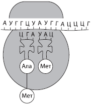
Each amino acid is encoded in DNA by three nucleotides. triplet for example, methionine is encoded by the TAC triplet, that is, the triplet code. On the other hand, each triplet encodes only one amino acid, which is its specificity or unambiguity. The genetic code is universal for all living organisms, that is, hereditary information about human proteins can be read by bacteria and vice versa. This testifies to the unity of the origin of the organic world. However, 64 combinations of three nucleotides correspond to only 20 amino acids, as a result of which 2-6 triplets can encode one amino acid, that is, the genetic code is redundant, or degenerate. Three triplets do not have corresponding amino acids, they are called stop codons, as they mark the end of the synthesis of the polypeptide chain.
The sequence of bases in DNA triplets and the amino acids they encode

*Stop codon, indicating the end of the synthesis of the polypeptide chain.
Abbreviations for amino acid names:
Ala - alanine
Arg - arginine
Asn - asparagine
Asp - aspartic acid
Val - valine
His - histidine
Gly - glycine
Gln - glutamine
Glu - glutamic acid
Ile - isoleucine
Leu - leucine
Liz - lysine
Meth - methionine
Pro - proline
Ser - serine
Tyr - tyrosine
Tre - threonine
Three - tryptophan
Fen - phenylalanine
cis - cysteine
If you start reading genetic information not from the first nucleotide in the triplet, but from the second, then not only will the reading frame shift, the protein synthesized in this way will be completely different not only in the nucleotide sequence, but also in structure and properties. There are no punctuation marks between the triplets, so there are no obstacles to the shift of the reading frame, which opens up scope for the occurrence and maintenance of mutations.
Matrix nature of biosynthetic reactions
Bacterial cells are able to duplicate every 20-30 minutes, and eukaryotic cells - every day and even more often, which requires high speed and accuracy of DNA replication. In addition, each cell contains hundreds and thousands of copies of many proteins, especially enzymes, therefore, for their reproduction, the "piece" method of their production is unacceptable. A more progressive way is stamping, which allows you to get numerous exact copies of the product and also reduce its cost. For stamping, a matrix is needed, with which an impression is made.
In cells, the principle of matrix synthesis is that new molecules of proteins and nucleic acids are synthesized in accordance with the program laid down in the structure of pre-existing molecules of the same nucleic acids (DNA or RNA).
Biosynthesis of protein and nucleic acids
DNA replication. DNA is a double-stranded biopolymer whose monomers are nucleotides. If DNA biosynthesis proceeded according to the principle of photocopying, then numerous distortions and errors in hereditary information would inevitably arise, which would ultimately lead to the death of new organisms. Therefore, the process of DNA duplication is different, in a semi-conservative way: the DNA molecule unwinds, and on each of the chains a new chain is synthesized according to the principle of complementarity. The process of self-reproduction of the DNA molecule, which ensures the exact copying of hereditary information and its transmission from generation to generation, is called replication(from lat. replication- repetition). As a result of replication, two absolutely exact copies of the parent DNA molecule are formed, each of which carries one copy of the parent.

The process of replication is actually extremely complex, since a number of proteins are involved in it. Some of them unwind the double helix of DNA, others break the hydrogen bonds between the nucleotides of complementary chains, others (for example, the DNA polymerase enzyme) select new nucleotides according to the principle of complementarity, etc. The two DNA molecules formed as a result of replication diverge in two during division. newly formed daughter cells.
Errors in the replication process are extremely rare, but if they do occur, they are very quickly eliminated by both DNA polymerases and special repair enzymes, since any error in the nucleotide sequence can lead to an irreversible change in the structure and functions of the protein and, ultimately, adversely affect the viability of a new cell or even an individual.
protein biosynthesis. As the outstanding philosopher of the 19th century F. Engels figuratively put it: "Life is a form of existence of protein bodies." The structure and properties of protein molecules are determined by their primary structure, i.e., the sequence of amino acids encoded in DNA. Not only the existence of the polypeptide itself, but also the functioning of the cell as a whole depends on the accuracy of reproduction of this information; therefore, the process of protein synthesis is of great importance. It seems to be the most complex process of synthesis in the cell, since up to three hundred different enzymes and other macromolecules are involved here. In addition, it flows at a high speed, which requires even greater precision.
There are two main steps in protein biosynthesis: transcription and translation.
Transcription(from lat. transcription- rewriting) is the biosynthesis of mRNA molecules on a DNA template.

Since the DNA molecule contains two antiparallel chains, reading information from both chains would lead to the formation of completely different mRNAs, therefore their biosynthesis is possible only on one of the chains, which is called coding, or codogenic, in contrast to the second, non-coding, or non-codogenic. The rewriting process is provided by a special enzyme, RNA polymerase, which selects RNA nucleotides according to the principle of complementarity. This process can take place both in the nucleus and in organelles that have their own DNA - mitochondria and plastids.
The mRNA molecules synthesized during transcription undergo a complex process of preparation for translation (mitochondrial and plastid mRNAs can remain inside organelles, where the second stage of protein biosynthesis takes place). In the process of mRNA maturation, the first three nucleotides (AUG) and a tail of adenyl nucleotides are attached to it, the length of which determines how many protein copies can be synthesized on a given molecule. Only then do mature mRNAs leave the nucleus through nuclear pores.
In parallel, the process of amino acid activation occurs in the cytoplasm, during which the amino acid is attached to the corresponding free tRNA. This process is catalyzed by a special enzyme, it consumes ATP.
Broadcast(from lat. broadcast- transfer) is the biosynthesis of a polypeptide chain on an mRNA template, in which genetic information is translated into a sequence of amino acids of the polypeptide chain.
The second stage of protein synthesis most often occurs in the cytoplasm, for example, on the rough endoplasmic reticulum. Its occurrence requires the presence of ribosomes, activation of tRNA, during which they attach the corresponding amino acids, the presence of Mg2+ ions, as well as optimal environmental conditions (temperature, pH, pressure, etc.).
To start broadcasting initiation) a small subunit of the ribosome is attached to the mRNA molecule ready for synthesis, and then, according to the principle of complementarity, the tRNA carrying the amino acid methionine is selected to the first codon (AUG). Only then does the large subunit of the ribosome join. Within the assembled ribosome, there are two mRNA codons, the first of which is already occupied. A second tRNA, also carrying an amino acid, is attached to the codon adjacent to it, after which a peptide bond is formed between the amino acid residues with the help of enzymes. The ribosome moves one codon of the mRNA; the first of the tRNA, freed from the amino acid, returns to the cytoplasm for the next amino acid, and a fragment of the future polypeptide chain, as it were, hangs on the remaining tRNA. The next tRNA joins the new codon, which is within the ribosome, the process repeats and step by step the polypeptide chain lengthens, i.e., it elongation.
End of protein synthesis termination) occurs as soon as a specific nucleotide sequence is encountered in an mRNA molecule that does not encode an amino acid (stop codon). After that, the ribosome, mRNA and polypeptide chain are separated, and the newly synthesized protein acquires the appropriate structure and is transported to the part of the cell where it will perform its functions.
Translation is a very energy-intensive process, since the energy of one ATP molecule is spent on attaching one amino acid to tRNA, and several more are used to move the ribosome along the mRNA molecule.
To accelerate the synthesis of certain protein molecules, several ribosomes can be sequentially attached to the mRNA molecule, which form a single structure - polysome.
The cell is the genetic unit of living things. Chromosomes, their structure (shape and size) and functions. The number of chromosomes and their species constancy. Somatic and sex cells. Cell life cycle: interphase and mitosis. Mitosis is the division of somatic cells. Meiosis. Phases of mitosis and meiosis. The development of germ cells in plants and animals. Cell division is the basis for the growth, development and reproduction of organisms. The role of meiosis and mitosis
The cell is the genetic unit of life
Despite the fact that nucleic acids are the carrier of genetic information, the implementation of this information is impossible outside the cell, which is easily proved by the example of viruses. These organisms, often containing only DNA or RNA, cannot reproduce on their own, for this they must use the hereditary apparatus of the cell. They cannot even penetrate the cell without the help of the cell itself, except by using the mechanisms of membrane transport or due to cell damage. Most viruses are unstable, they die after a few hours of exposure to the open air. Therefore, the cell is a genetic unit of the living, which has a minimum set of components for the preservation, modification and implementation of hereditary information, as well as its transmission to descendants.
Most of the genetic information of a eukaryotic cell is located in the nucleus. A feature of its organization is that, unlike the DNA of a prokaryotic cell, eukaryotic DNA molecules are not closed and form complex complexes with proteins - chromosomes.
Chromosomes, their structure (shape and size) and functions
Chromosome(from Greek. chrome- color, color and catfish- body) is the structure of the cell nucleus, which contains genes and carries certain hereditary information about the signs and properties of the organism.
Sometimes the ring DNA molecules of prokaryotes are also called chromosomes. Chromosomes are capable of self-duplication, they have a structural and functional individuality and retain it in a number of generations. Each cell carries all the hereditary information of the body, but only a small part of it works.
The basis of the chromosome is a double-stranded DNA molecule packed with proteins. In eukaryotes, histone and non-histone proteins interact with DNA, while in prokaryotes, histone proteins are absent.
Chromosomes are best seen under a light microscope during cell division, when, as a result of compaction, they take the form of rod-shaped bodies separated by a primary constriction - centromere — on shoulders. The chromosome may also have secondary constriction, which in some cases separates the so-called satellite. The ends of chromosomes are called telomeres. Telomeres prevent the ends of chromosomes from sticking together and ensure their attachment to the nuclear membrane in a non-dividing cell. At the beginning of division, the chromosomes are doubled and consist of two daughter chromosomes - chromatids attached at the centromere.

According to the shape, equal-arm, unequal-arm and rod-shaped chromosomes are distinguished. Chromosome sizes vary significantly, but the average chromosome has a size of 5 $×$ 1.4 µm.
In some cases, chromosomes, as a result of numerous DNA duplications, contain hundreds and thousands of chromatids: such giant chromosomes are called polythene. They are found in the salivary glands of Drosophila larvae, as well as in the digestive glands of roundworms.
The number of chromosomes and their species constancy. Somatic and germ cells
According to the cellular theory, a cell is a unit of structure, life and development of an organism. Thus, such important functions of living things as growth, reproduction and development of the organism are provided at the cellular level. Cells of multicellular organisms can be divided into somatic and sex.
somatic cells are all the cells of the body that are formed as a result of mitotic division.
The study of chromosomes made it possible to establish that the somatic cells of the organism of each biological species are characterized by a constant number of chromosomes. For example, a person has 46 of them. The set of chromosomes of somatic cells is called diploid(2n), or double.

sex cells, or gametes, are specialized cells that serve for sexual reproduction.
Gametes always contain half as many chromosomes as in somatic cells (in humans - 23), so the set of chromosomes of germ cells is called haploid(n), or single. Its formation is associated with meiotic cell division.
The amount of DNA of somatic cells is designated as 2c, and that of germ cells is 1c. The genetic formula of somatic cells is written as 2n2c, and sex - 1n1c.
In the nuclei of some somatic cells, the number of chromosomes may differ from their number in somatic cells. If this difference is greater by one, two, three, etc. haploid sets, then such cells are called polyploid(tri-, tetra-, pentaploid, respectively). In such cells, metabolic processes are usually very intensive.
The number of chromosomes in itself is not a species-specific trait, since different organisms may have the same number of chromosomes, while related ones may have different numbers. For example, malarial plasmodium and horse roundworm have two chromosomes, while humans and chimpanzees have 46 and 48, respectively.
Human chromosomes are divided into two groups: autosomes and sex chromosomes (heterochromosomes). Autosome there are 22 pairs in human somatic cells, they are the same for men and women, and sex chromosomes only one pair, but it is she who determines the sex of the individual. There are two types of sex chromosomes - X and Y. The cells of the body of a woman carry two X chromosomes, and men - X and Y.
Karyotype- this is a set of signs of the chromosome set of an organism (the number of chromosomes, their shape and size).
The conditional record of the karyotype includes the total number of chromosomes, sex chromosomes, and possible deviations in the set of chromosomes. For example, the karyotype of a normal male is written as 46,XY, while the karyotype of a normal woman is 46,XX.
Cell life cycle: interphase and mitosis
Cells do not arise each time anew, they are formed only as a result of the division of mother cells. After separation, the daughter cells take some time to form organelles and acquire the appropriate structure that would ensure the performance of a certain function. This period of time is called ripening.
The period of time from the appearance of a cell as a result of division to its division or death is called cell life cycle.
In eukaryotic cells, the life cycle is divided into two main stages: interphase and mitosis.
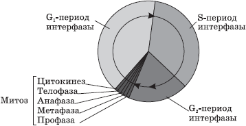
Interphase- this is the period of time in the life cycle in which the cell does not divide and functions normally. The interphase is divided into three periods: G 1 -, S- and G 2 -periods.
G 1 -period(presynthetic, postmitotic) is a period of cell growth and development, during which there is an active synthesis of RNA, proteins and other substances necessary for the complete life support of the newly formed cell. By the end of this period, the cell may begin to prepare for DNA duplication.
AT S-period(synthetic) the process of DNA replication takes place. The only part of the chromosome that does not undergo replication is the centromere, therefore, the resulting DNA molecules do not completely diverge, but remain fastened in it, and at the beginning of division, the chromosome has an X-shaped appearance. The genetic formula of the cell after DNA duplication is 2n4c. Also in the S-period, doubling of the centrioles of the cell center occurs.
G 2 -period(postsynthetic, premitotic) is characterized by intensive synthesis of RNA, proteins and ATP necessary for the process of cell division, as well as the separation of centrioles, mitochondria and plastids. Until the end of the interphase, the chromatin and nucleolus remain clearly distinguishable, the integrity of the nuclear membrane is not disturbed, and the organelles do not change.
Some of the cells of the body are able to perform their functions throughout the life of the body (neurons of our brain, muscle cells of the heart), while others exist for a short time, after which they die (cells of the intestinal epithelium, cells of the epidermis of the skin). Consequently, the processes of cell division and the formation of new cells must constantly occur in the body, which would replace the dead ones. Cells capable of dividing are called stem. In the human body, they are found in the red bone marrow, in the deep layers of the epidermis of the skin and other places. Using these cells, you can grow a new organ, achieve rejuvenation, and also clone the body. The prospects for the use of stem cells are quite clear, but the moral and ethical aspects of this problem are still being discussed, since in most cases embryonic stem cells obtained from human embryos killed during abortion are used.
The duration of interphase in plant and animal cells averages 10-20 hours, while mitosis takes about 1-2 hours.
In the course of successive divisions in multicellular organisms, daughter cells become more and more diverse, as they read information from an increasing number of genes.
Some cells eventually stop dividing and die, which may be due to the completion of certain functions, as in the case of epidermal cells of the skin and blood cells, or to damage to these cells by environmental factors, in particular pathogens. Genetically programmed cell death is called apoptosis while accidental death is necrosis.
Mitosis is the division of somatic cells. Phases of mitosis
Mitosis- a method of indirect division of somatic cells.
During mitosis, the cell goes through a series of successive phases, as a result of which each daughter cell receives the same set of chromosomes as in the mother cell.
Mitosis is divided into four main phases: prophase, metaphase, anaphase, and telophase. Prophase- the longest stage of mitosis, during which chromatin condensation occurs, as a result of which X-shaped chromosomes, consisting of two chromatids (daughter chromosomes), become visible. In this case, the nucleolus disappears, the centrioles diverge towards the poles of the cell, and the achromatin spindle (spindle) of microtubules begins to form. At the end of prophase, the nuclear membrane breaks up into separate vesicles.

AT metaphase chromosomes line up along the equator of the cell with their centromeres, to which microtubules of a fully formed division spindle are attached. At this stage of division, the chromosomes are most dense and have a characteristic shape, which makes it possible to study the karyotype.
AT anaphase rapid DNA replication occurs in the centromeres, as a result of which the chromosomes split and the chromatids diverge towards the poles of the cell, stretched by microtubules. The distribution of chromatids must be absolutely equal, since it is this process that maintains the constancy of the number of chromosomes in the cells of the body.
On the stage telophase daughter chromosomes gather at the poles, despiralize, around them nuclear envelopes form from the vesicles, and nucleoli appear in the newly formed nuclei.
After the division of the nucleus, the division of the cytoplasm occurs - cytokinesis, during which there is a more or less uniform distribution of all the organelles of the mother cell.
Thus, as a result of mitosis, two daughter cells are formed from one mother cell, each of which is a genetic copy of the mother cell (2n2c).
In diseased, damaged, aging cells and specialized tissues of the body, a slightly different division process can occur - amitosis. Amitosis called direct division of eukaryotic cells, in which the formation of genetically equivalent cells does not occur, since the cellular components are distributed unevenly. It occurs in plants in the endosperm and in animals in the liver, cartilage, and cornea of the eye.
Meiosis. Phases of meiosis
Meiosis- this is a method of indirect division of primary germ cells (2n2c), as a result of which haploid cells (1n1c), most often germ cells, are formed.
Unlike mitosis, meiosis consists of two successive cell divisions, each preceded by an interphase. The first division of meiosis (meiosis I) is called reduction, since in this case the number of chromosomes is halved, and the second division (meiosis II) - equational, since in its process the number of chromosomes is conserved.

Interphase I proceeds similarly to the interphase of mitosis. Meiosis I is divided into four phases: prophase I, metaphase I, anaphase I and telophase I. prophase I Two major processes occur: conjugation and crossing over. Conjugation- this is the process of fusion of homologous (paired) chromosomes along the entire length. The pairs of chromosomes formed during conjugation are retained until the end of metaphase I.
Crossing over- mutual exchange of homologous regions of homologous chromosomes. As a result of crossing over, the chromosomes received by the organism from both parents acquire new combinations of genes, which leads to the appearance of genetically diverse offspring. At the end of prophase I, as in the prophase of mitosis, the nucleolus disappears, the centrioles diverge towards the poles of the cell, and the nuclear envelope disintegrates.

AT metaphase I pairs of chromosomes line up along the equator of the cell, microtubules of the fission spindle are attached to their centromeres.
AT anaphase I whole homologous chromosomes consisting of two chromatids diverge to the poles.
AT telophase I around clusters of chromosomes at the poles of the cell, nuclear membranes form, nucleoli form.
Cytokinesis I provides division of cytoplasms of daughter cells.
The daughter cells formed as a result of meiosis I (1n2c) are genetically heterogeneous, since their chromosomes, randomly dispersed to the poles of the cell, contain unequal genes.
Comparative characteristics of mitosis and meiosis
| sign | Mitosis | Meiosis | |
| What cells start dividing? | Somatic (2n) | Primary germ cells (2n) | |
| Number of divisions | 1 | 2 | |
| How many and what kind of cells are formed in the process of division? | 2 somatic (2n) | 4 sexual (n) | |
| Interphase | Cell preparation for division, DNA duplication | Very short, DNA duplication does not occur | |
| Phases | Meiosis I | Meiosis II | |
| Prophase | Chromosome condensation, disappearance of the nucleolus, disintegration of the nuclear envelope, conjugation and crossing over may occur | Condensation of chromosomes, disappearance of the nucleolus, disintegration of the nuclear envelope | |
| metaphase | Pairs of chromosomes are located along the equator, a division spindle is formed | Chromosomes line up along the equator, the spindle of division is formed | |
| Anaphase | Homologous chromosomes from two chromatids diverge towards the poles | Chromatids diverge towards the poles | |
| Telophase | Chromosomes despiralize, new nuclear envelopes and nucleoli form | Chromosomes despiralize, new nuclear envelopes and nucleoli form | |
Interphase II very short, since DNA doubling does not occur in it, that is, there is no S-period.
Meiosis II also divided into four phases: prophase II, metaphase II, anaphase II and telophase II. AT prophase II the same processes occur as in prophase I, with the exception of conjugation and crossing over.
AT metaphase II Chromosomes are located along the equator of the cell.
AT anaphase II Chromosomes split at the centromere and the chromatids stretch towards the poles.
AT telophase II nuclear membranes and nucleoli form around clusters of daughter chromosomes.
After cytokinesis II the genetic formula of all four daughter cells is 1n1c, but they all have a different set of genes, which is the result of crossing over and a random combination of maternal and paternal chromosomes in daughter cells.
The development of germ cells in plants and animals
Gametogenesis(from Greek. gamete- wife, gametes- husband and genesis- origin, occurrence) is the process of formation of mature germ cells.
Since sexual reproduction most often requires two individuals - female and male, producing different sex cells - eggs and sperm, then the processes of formation of these gametes should be different.
The nature of the process also depends to a large extent on whether it occurs in a plant or animal cell, since in plants only mitosis occurs during the formation of gametes, while in animals both mitosis and meiosis occur.
The development of germ cells in plants. In angiosperms, the formation of male and female germ cells occurs in different parts of the flower - stamens and pistils, respectively.
Before the formation of male germ cells - microgametogenesis(from Greek. micros- small) - happening microsporogenesis, that is, the formation of microspores in the anthers of the stamens. This process is associated with the meiotic division of the mother cell, which results in four haploid microspores. Microgametogenesis is associated with the mitotic division of microspores, giving a male gametophyte of two cells - a large vegetative(siphonogenic) and shallow generative. After division, the male gametophyte is covered with dense shells and forms a pollen grain. In some cases, even in the process of pollen maturation, and sometimes only after transfer to the stigma of the pistil, the generative cell divides mitotically with the formation of two immobile male germ cells - sperm. After pollination, a pollen tube is formed from the vegetative cell, through which sperm penetrate into the ovary of the pistil for fertilization.

The development of female germ cells in plants is called megagametogenesis(from Greek. megas- big). It occurs in the ovary of the pistil, which is preceded by megasporogenesis, as a result of which four megaspores are formed from the mother cell of the megaspore lying in the nucellus by meiotic division. One of the megaspores divides mitotically three times, giving rise to the female gametophyte, an embryo sac with eight nuclei. With the subsequent isolation of the cytoplasms of the daughter cells, one of the resulting cells becomes an egg, on the sides of which lie the so-called synergids, three antipodes are formed at the opposite end of the embryo sac, and in the center, as a result of the fusion of two haploid nuclei, a diploid central cell is formed.

The development of germ cells in animals. In animals, two processes of the formation of germ cells are distinguished - spermatogenesis and oogenesis.
spermatogenesis(from Greek. sperm, spermatos- seed and genesis- origin, occurrence) is the process of formation of mature male germ cells - spermatozoa. In humans, it occurs in the testes, or testes, and is divided into four periods: reproduction, growth, maturation and formation.

AT breeding season primordial germ cells divide mitotically, resulting in the formation of diploid spermatogonia. AT growth period spermatogonia accumulate nutrients in the cytoplasm, increase in size and turn into primary spermatocytes, or spermatocytes of the 1st order. Only after that they enter meiosis ( ripening period), which first results in two secondary spermatocyte, or spermatocyte of the 2nd order, and then - four haploid cells with a fairly large amount of cytoplasm - spermatids. AT formation period they lose almost all of the cytoplasm and form a flagellum, turning into spermatozoa.
spermatozoa, or gummies, - very small mobile male sex cells with a head, neck and tail.
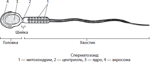
AT head, except for the core, is acrosome- a modified Golgi complex, which ensures the dissolution of the membranes of the egg during fertilization. AT neck there are centrioles of the cell center, and the basis ponytail form microtubules that directly support the movement of the spermatozoon. It also contains mitochondria, which provide the sperm with ATP energy for movement.
Ovogenesis(from Greek. UN- an egg and genesis- origin, occurrence) is the process of formation of mature female germ cells - eggs. In humans, it occurs in the ovaries and consists of three periods: reproduction, growth and maturation. Periods of reproduction and growth, similar to those in spermatogenesis, occur even during intrauterine development. At the same time, diploid cells are formed from the primary germ cells as a result of mitosis. oogonia, which then turn into diploid primary oocytes, or oocytes of the 1st order. Meiosis and subsequent cytokinesis occurring in ripening period, are characterized by uneven division of the cytoplasm of the mother cell, so that as a result, at first one is obtained secondary oocyte, or oocyte 2nd order, and first polar body, and then from the secondary oocyte - the egg, which retains the entire supply of nutrients, and the second polar body, while the first polar body is divided into two. Polar bodies take away excess genetic material.
In humans, eggs are produced with an interval of 28-29 days. The cycle associated with the maturation and release of eggs is called the menstrual cycle.
Egg- a large female germ cell, which carries not only a haploid set of chromosomes, but also a significant supply of nutrients for the subsequent development of the embryo.

The egg in mammals is covered with four membranes, which reduce the likelihood of damage to it by various factors. The diameter of the egg in humans reaches 150-200 microns, while in an ostrich it can be several centimeters.
Cell division is the basis for the growth, development and reproduction of organisms. The role of mitosis and meiosis
If in unicellular organisms cell division leads to an increase in the number of individuals, i.e., reproduction, then in multicellular organisms this process can have a different meaning. Thus, cell division of the embryo, starting from the zygote, is the biological basis for the interconnected processes of growth and development. Similar changes are observed in a person during adolescence, when the number of cells not only increases, but also a qualitative change in the body occurs. Reproduction of multicellular organisms is also based on cell division, for example, during asexual reproduction, due to this process, a whole body is restored from a part of the organism, and during sexual reproduction, germ cells are formed during gametogenesis, subsequently giving a new organism. It should be noted that the main methods of eukaryotic cell division—mitosis and meiosis—have different significance in the life cycles of organisms.
As a result of mitosis, there is a uniform distribution of hereditary material between daughter cells - exact copies of the mother. Without mitosis, the existence and growth of multicellular organisms developing from a single cell, the zygote, would be impossible, since all cells of such organisms must contain the same genetic information.
In the process of division, daughter cells become more and more diverse in structure and functions, which is associated with the activation of new groups of genes in them due to intercellular interaction. Thus, mitosis is necessary for the development of an organism.
This method of cell division is necessary for the processes of asexual reproduction and regeneration (recovery) of damaged tissues, as well as organs.
Meiosis, in turn, ensures the constancy of the karyotype during sexual reproduction, as it reduces by half the set of chromosomes before sexual reproduction, which is then restored as a result of fertilization. In addition, meiosis leads to the appearance of new combinations of parental genes due to crossing over and random combination of chromosomes in daughter cells. Thanks to this, the offspring is genetically diverse, which provides material for natural selection and is the material basis of evolution. A change in the number, shape and size of chromosomes, on the one hand, can lead to the appearance of various deviations in the development of the organism and even its death, and on the other hand, it can lead to the appearance of individuals more adapted to the environment.
Thus, the cell is a unit of growth, development and reproduction of organisms.
CELL
EPITHELIAL TISSUE.
TYPES OF FABRICS.
STRUCTURE AND PROPERTIES OF THE CELL.
LECTURE №2.
1. The structure and basic properties of the cell.
2. The concept of tissues. Types of fabrics.
3. Structure and functions of epithelial tissue.
4. Types of epithelium.
Purpose: to know the structure and properties of the cell, types of tissues. Present the classification of the epithelium and its location in the body. To be able to distinguish epithelial tissue by morphological features from other tissues.
1. A cell is an elementary living system, the basis of the structure, development and life of all animals and plants. The science of the cell is cytology (Greek cytos - cell, logos - science). Zoologist T. Schwann in 1839 for the first time formulated the cellular theory: the cell is the basic structural unit of all living organisms, the cells of animals and plants are similar in structure, there is no life outside the cell. Cells exist as independent organisms (protozoa, bacteria), and as part of multicellular organisms, in which there are sex cells that serve for reproduction, and body cells (somatic), different in structure and functions (nerve, bone, secretory, etc.). ).The sizes of human cells range from 7 microns (lymphocytes) to 200-500 microns (female egg, smooth myocytes). Any cell contains proteins, fats, carbohydrates, nucleic acids, ATP, mineral salts and water. From inorganic substances, the cell contains the most water (70-80%), from organic - proteins (10-20%). The main parts of the cell are: nucleus, cytoplasm, cell membrane (cytolemma).
NUCLEUS CYTOPLASMA CYTOLEMMA
Nucleoplasm - hyaloplasm
1-2 nucleoli - organelles
Chromatin (endoplasmic reticulum)
complex Ktolji
cell center
mitochondria
lysosomes
special purpose)
Inclusions.
The nucleus of the cell is located in the cytoplasm and is separated from it by the nuclear
shell - nucleolemma. It serves as a site for genes
the main chemical substance of which is DNA. The nucleus regulates the shaping processes of the cell and all its vital functions. Nucleoplasm ensures the interaction of various nuclear structures, nucleoli are involved in the synthesis of cellular proteins and some enzymes, chromatin contains chromosomes with genes that carry heredity.
Hyaloplasm (Greek hyalos - glass) - the main plasma of the cytoplasm,
is the true internal environment of the cell. It unites all cellular ultrastructures (nucleus, organelles, inclusions) and ensures their chemical interaction with each other.
Organelles (organelles) are permanent ultrastructures of the cytoplasm that perform certain functions in the cell. These include:
1) endoplasmic reticulum - a system of branched channels and cavities formed by double membranes associated with the cell membrane. On the walls of the channels there are tiny little bodies - ribosomes, which are centers of protein synthesis;
2) the K. Golgi complex, or the internal mesh apparatus, has meshes and contains vacuoles of various sizes (lat. Vacuum - empty), participates in the excretory function of cells and in the formation of lysosomes;
3) the cell center - the cytocenter consists of a spherical dense body - the centrosphere, inside which lie 2 dense bodies - centrioles, interconnected by a bridge. It is located closer to the nucleus, takes part in cell division, ensuring an even distribution of chromosomes between daughter cells;
4) mitochondria (Greek mitos - thread, chondros - grain) look like grains, sticks, threads. They carry out the synthesis of ATP.
5) lysosomes - vesicles filled with enzymes that regulate
metabolic processes in the cell and have digestive (phagocytic) activity.
6) special-purpose organelles: myofibrils, neurofibrils, tonofibrils, cilia, villi, flagella, performing a specific cell function.
Cytoplasmic inclusions are non-permanent formations in the form
granules, drops and vacuoles containing proteins, fats, carbohydrates, pigment.
The cell membrane - the cytolemma, or plasmolemma, covers the cell from the surface and separates it from the environment. It is semi-permeable and regulates the entry of substances into the cell and their exit from it.
The intercellular substance is located between cells. In some tissues, it is liquid (for example, in the blood), while in others it consists of an amorphous (structureless) substance.
Any living cell has the following basic properties:
1) metabolism, or metabolism (the main vital property),
2) sensitivity (irritability);
3) the ability to reproduce (self-reproduce);
4) the ability to grow, i.e. an increase in the size and volume of cellular structures and the cell itself;
5) the ability to develop, i.e. the acquisition by the cell of specific functions;
6) secretion, i.e. release of various substances;
7) movement (leukocytes, histiocytes, spermatozoa)
8) phagocytosis (leukocytes, macrophages, etc.).
2. Tissue is a system of cells similar in origin), structure and functions. The composition of tissues also includes tissue fluid and waste products of cells. The doctrine of tissues is called histology (Greek histos - tissue, logos - teaching, science). In accordance with the characteristics of the structure, function and development, the following types of tissues are distinguished:
1) epithelial, or integumentary;
2) connective (tissues of the internal environment);
3) muscular;
4) nervous.
A special place in the human body is occupied by blood and lymph - a liquid tissue that performs respiratory, trophic and protective functions.
In the body, all tissues are closely related morphologically.
and functional. The morphological connection is due to the fact that different
nye tissues are part of the same organs. functional connection
is manifested in the fact that the activity of different tissues that make up the
bodies, agreed.
Cellular and non-cellular elements of tissues in the process of life
activities wear out and die (physiological degeneration)
and recover (physiological regeneration). When damaged
tissues are also restored (reparative regeneration).
However, this process is not the same for all tissues. Epithelial
naya, connective, smooth muscle tissue and blood cells regenerate
roar well. striated muscle tissue is restored
only under certain conditions. are restored in the nervous tissue
only nerve fibers. The division of nerve cells in the body of an adult
person has not been identified.
3. Epithelial tissue (epithelium) is a tissue that covers the surface of the skin, the cornea of the eye, and also lines all the cavities of the body, the inner surface of the hollow organs of the digestive, respiratory, genitourinary systems, is part of most of the body's glands. In this regard, there are integumentary and glandular epithelium.
The integumentary epithelium, being the border tissue, carries out:
1) a protective function, protecting the underlying tissues from various external influences: chemical, mechanical, infectious.
2) the body's metabolism with the environment, performing the functions of gas exchange in the lungs, absorption in the small intestine, excretion of metabolic products (metabolites);
3) creation of conditions for the mobility of internal organs in serous cavities: heart, lungs, intestines, etc.
The glandular epithelium performs a secretory function, i.e. it forms and secretes specific products - secrets that are used in the processes occurring in the body.
Morphologically, epithelial tissue differs from other body tissues in the following ways:
1) it always occupies a borderline position, since it is located on the border of the external and internal environments of the body;
2) it is a layer of cells - epitheliocytes, which have an unequal shape and structure in different types of epithelium;
3) there is no intercellular substance between the epithelial cells, and the cells
connected to each other through various contacts.
4) epithelial cells are located on the basal membrane (a plate about 1 micron thick, by which it is separated from the underlying connective tissue. The basal membrane consists of an amorphous substance and fibrillar structures;
5) epithelial cells have polarity, i.e. the basal and apical sections of the cells have a different structure;
6) the epithelium does not contain blood vessels, so cell nutrition
carried out by diffusion of nutrients through the basement membrane from the underlying tissues;
7) the presence of tonofibrils - filamentous structures that give strength to epithelial cells.
4. There are several classifications of the epithelium, which are based on various features: origin, structure, functions. Of these, the most widespread is the morphological classification, taking into account the relationship of cells to the basement membrane and their shape on the free apical (Latin apex - top) part of the epithelial layer . This classification reflects the structure of the epithelium, depending on its function.
Single-layer squamous epithelium is represented in the body by endothelium and mesothelium. The endothelium lines the blood vessels, lymphatic vessels, and chambers of the heart. The mesothelium covers the serous membranes of the peritoneal cavity, pleura and pericardium. A single layer of cuboidal epithelium lines part of the renal tubules, ducts of many glands, and small bronchi. A single-layer prismatic epithelium has a mucous membrane of the stomach, small and large intestines, uterus, fallopian tubes, gallbladder, a number of ducts of the liver, pancreas, part
kidney tubules. In organs where absorption processes occur, epithelial cells have a suction border consisting of a large number of microvilli. A single-layer multi-row ciliated epithelium lines the airways: the nasal cavity, nasopharynx, larynx, trachea, bronchi, etc.
The stratified squamous non-keratinized epithelium covers the outside of the cornea of the eye and the mucous membrane of the oral cavity and esophagus. The stratified squamous keratinized epithelium forms the surface layer of the cornea and is called the epidermis. The transitional epithelium is typical for urinary organs: renal pelvis, ureters, bladder, the walls of which are subject to significant stretching when filled with urine.
Exocrine glands secrete their secret into the cavity of the internal organs or onto the surface of the body. They usually have excretory ducts. Endocrine glands do not have ducts and secrete secretions (hormones) into the blood or lymph.